Mastering the Fetal Great Vessels - Part 7-1
The 3 Vessel View
The 3 Vessel View
what is the 3-vessel view
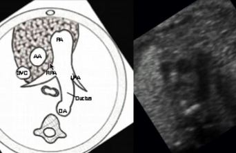
- The 3 vessel view is essentially an axial image through the great vessels, You will see:
- The pulmonary artery is seen as the most lateral vessel and will “continue" into (either) the ductus arteriosus or branch into the left or right pulmonary artery
- The aorta in cross-section
- The superior vena cava in cross-section
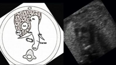
3 key observations to make on the 3 vessel view
- The vessel on the end is branching (which makes this the pulmonary artery). This could be the left or right pulmonary artery or the ductus arteriosus
- The pulmonary artery is anterior to the aorta
- The pulmonary artery and aorta are about the same size
- Extra Information: The right pulmonary artery travels behind and touches the aorta
superior vena cava
- Most of the focus of the 3-vessel view is on the pulmonary artery and aorta, however, let's not forget that "third vessel"
- Make a point to look at the SVC and make an assessment whether it is too small or too big.
- If either is the case, recommend a fetal echo as these findings are associated with other possible anomalies
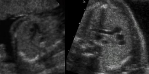
what if we see "4 vessels" on the "3 vessel" view
- If you see a 4th vessel in cross-section adjacent to the pulmonary artery, this is, most commonly, a persistent left SVC
- While this entity itself is not specifically dangerous, it can be associated with other anomalies, so......
- You guessed it! RECOMMEND A FETAL ECHO