
Readout - How to determine if cyst is paraovarian or exophytic ovarian (and what to do with it)
Take Home Points
clues to cyst being paraovarian vs. exophytic ovarian
- Compression: This is a technique we use frequently.
- Using the endovaginal transducer, place the tip between the two structures that you want to evaluate and use pressure on the probe.
- If the structures move together, they are connected and if they spread apart, you can infer that they are separate structures.
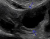
- In this video, had the cyst separated from the ovary, we would have know that it was a distinct paraovarian cyst.
- Claw sign: When the ovarian parenchyma forms a "claw" of tissue partially surrounds the cyst, it is intraovarian
is followup necessary?
- Paraovarian cysts (which this was not) are generally innocuous, but not entirely.
- While the pathogenesis of paraovarian cysts is different from ovarian cysts, the Society of Radiologist in Ultrasound decided to apply the same followup recommendations to both types of lesions.
official sru statement
- Paraovarian and paratubal cysts were considered together with ovarian cysts.
- Unlike the ovary, where folliculogenesis usually explains follicles up to 3 cm, we recognize that there is no similar rationale for ignoring small simple paraovarian cysts.
- However, paraovarian cysts are common and usually appear sonographically as simple cysts. Simple paraovarian cysts are very unlikely to be malignant.
- While they are not likely to resolve, simple paraovarian cysts generally are inconsequential in asymptomatic women.
- The panel felt that using the same size thresholds as for ovarian cysts was reasonable.
- Adapted from: Management of Asymptomatic Ovarian and Other Adnexal Cysts Imaged at US: Society of Radiologists in Ultrasound Consensus Conference Statement