Nuchal Thickness Measurement
Tips, Tricks and Significance
Tips, Tricks and Significance
Take Home Points
what is the importance of the nuchal thickness measurement
- First, nuchal fold thickness measurement (typically performed between 16-20 weeks) must be differentiated from the nuchal translucency measurement done in the first trimester.
- Unlike other "soft markers" for chromosome abnormalities such as echogenic intracardiac foci, mild pyelectasis and choroid plexus cysts, thickening of the nuchal fold is a much "harder soft marker."
- A thickened nuchal fold increases the risk of Down's Syndrome by 17 times.
- In our opinion, every fetus with true nuchal fold thickening should undergo chromosomal analysis.
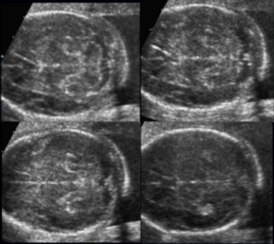
What are the landmarks for measuring the nuchal fold (axial plane)
- Cavum septi pellucidi
- 3rd ventricle (thalami)
- Cerebellum
- Cisterna Magna
- Occipital Bone
Pitfalls
- Measuring in the oblique axial plane has some definitive technical issues.
- Easy to spuriously increase the measurement
- Particularly if you scan with too much obliquity through the foramen magnum
- Particularly if you scan with too much obliquity through the foramen magnum
- The occipital bone is often shadowed out in this image
- This is due to the critical angle of reflection artifact
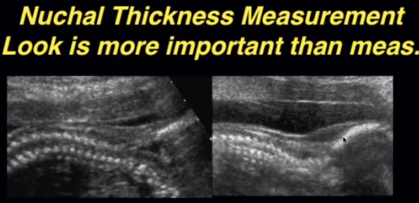
MID-LINE sagittal plane measurement
- When the nuchal fold measures too thick in the axial plane or we are having trouble getting an accurate measurement, we always confirm the measurement in the mid-sagittal plane.
- Measurement should be done it the very inferior aspect of the occipital bone, perpendicular to the bone.
- The mid-sagittal plane also has a pitfall. The fetus should have it's neck in neutral position. if the neck is extended, it will spuriously increase the measurement.
WHAT IS Normal measurement FOR NUCHAL THICKNESS
- Some use < 5 mm as the normal measurement up to 18 weeks and then < 6 mm from 18-29 weeks.
- Some use nuchal thickness up to 22-24 weeks, but we don't do that
- Some use < 5 mm as the normal measurement up to 18 weeks.


